A collection of complex joint preservation and replacement case studies and random thoughts of a orthopedic surgeon essentially aimed at knowledge dissemination.
Sunday, 25 July 2010
Arthrokochi
Tuesday, 6 July 2010

Friday, 11 June 2010
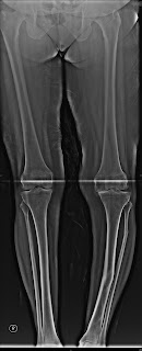
Tibia Vara Causing OA knee

Planned to do a metaphyseal corrective osteotomy followed by a TKR- one stage and grafting. Touch wt bearing for 6 weeks. Fibula was not osteotomised as adequete correction and compression and grafting was thought to be sufficient. 

 79 year 10 years post acetabular fracture fixed elsewhere presented with Trochanteric fracture. Patient ASA grade 2, non diabetic and pretty active. The judet views showed adequate posterior column and wall. Planned for a THR. Cemented or uncemented. 36 head if possible.
We decided to remove the implant only if it was interfereing and finally went thru modified hardinge and did a cemented repalcement. We wanted a larger head diameter with an uncemented cup but due to the presence of 2 intraarticular screw we cemented the same and a calcar replacement stem with wiring of the trochanter.

Thursday, 29 April 2010


68 year old patient with metaphyseal varus deformity of 22 degrees . Planned for TKR.
Friday, 5 March 2010


Comments please
In

 view of his age and communition we planned
  ORIF
ORIF  and primary THR. His haemoglobin was 8 gms we made us think of 1. fixing the posterior column and wall and use a multiholed cup if good,stable posterior superior host bone contact is obtained or 2.cage if we cant get the same.
attached are the pics with good posterior superior contact and primary THR. Host bone autografy used medialy to avoid protrusio.
 On your rt is the 3 month xray and is now full wt bearing> no migration detected yet.
Tuesday, 15 December 2009
54 year DDH


54 year DDH with pain rt. hip, Gross trendelenberg gait and 7 cm shortening.
Options
1. Milch Batchelor osteotomy if the patient is financially challenged to avoid trendelenberg gait and excision of the head if one believes that as a cause of pain-
2. THR with a modular stem with subtrochanteric shortening
Problems with THR- Solutions
Finding the true acetabulum- Standard hardinge exept the proximal limb in an acute angle to follow the gluteus medius fibres. Neck osteotomy as planned. Follow the inferior capsule from lesser trochanter to reach the true acetabulum or walk on the ilium with a Homan distally till you slip below the transverse ligament, all the while excising the scar.
acetabulum with narrower AP diameter as compared to supero inferior diameter.Thin or deficient anterior wall.- Drill the medial wall eith a 2.5 drill and measure the depth usually 1 to 1.5 cm. That is how much you can medialise the cup or even break the medial wall as described by Zhiang. Use the next reamers in the inclination and version decided with a posterior vector to ream less of the anterior wall till one gets a good fit not just superior inferior fit. 70% host bone contact is achieved without much ado.
Osteoporotic acetabulum as it was never loaded - .I had a problem in one case when the acetabular dome screws cut out when mobilising the patient and had to cement a cup. So keep Cemented cup back up.
Shortening of femur - well descibed techniques in literature- shall elucidate if needed without boring others with this writen diarrhoea.
Narrow canal
Proximal distal mismatch
2 yr follow up xrays after the weekend
As a reply to Dr. Utkarsh comments. To find the true acetabulum after the osteotomy force the spike of the homan;s spike on the ilium and excise the scar. Walk down on the ilium with the spike feeling the bone till you come to the deficiency inferiorly which is the tear drop.
As far as the stem goes I use a modular stem which is distal fixing and proximal loading with sizes upto 6m and small offset to cover for the narrow canal. The distal slot helps to hold the distal fragment after the shortening osteotomy and rarely a unicortical plate is needed.
There ia a latin american paper where in a distal shorterning osteotomy is in the metaphysis ( supracondylar)with similar results. Others have desribed a method to Intesucept the distal smaller dia fragment to the larger dia proximal fragment for stability. We use a sagital osteotomy of the shortenend excised fragments as a vascularised graft with V. lateralis attached to lie on either side of the osteotomy as seen in the above xrays.
.
perprosthetic fracture in a octogenarian-


perioprosthetic fracture in an 89 yr old male after Austin moore prosthesis implanted 3 years ago. COPD, H/0 recent CVA.
Ideally I want an implant which is cemented distallt and uncemented proximally for fractur healing and immediate mobilisation in view of his age. What was done was a fully porocoated cobalt chrome implant here in the porocoat was removed with a burr smoothen the stem for cementing distally and wiring proximally in the uncemented part. Patient was mobilised immediated post op with a walker wt bearing as comfortable. He is 8 months postop so far with no lysis. bipolar head used as the acetabulum was normal and low demand.
Hey I did a Restoration fluted stem revision yesterday- which may be an option here as it is titanium. fluted with grooves like wagner to fit distally and HA coated proximally for this octogenarian for immediate wt. bearing.
Saturday, 5 December 2009
To fix or replace -5 month old fracture dislocation


Sunday, 29 November 2009
Arthroscopic shoulder stabilisation.
Monday, 16 November 2009
Ankylosed knee with patellar fracture - by Sreenath for opinion
 54 yr male with trauma history of ankylosis same knee following septic arthritis at 16 yrs of age,diabetes controlled by dietnot willing for tkr as he had thought about it becos of ankylosis earlier and firm on that decision,no pain previously,office job
 Questions for academic interest
OATS

Similar defect in the lateral femoral condyle covered by a bioscaphold( Trufit) in a 40 year old man with equally good short term result-18 months
Friday, 13 November 2009
arthroscopic decompression of Spinoglenoid ganglion


25 yr old male, Pain ,weakness -7 months insiduous onset
No history of injury. Conservative treatment for 6 months elsewhere
external rotation weakness (rt) shoulder, Wasting of infraspinatus
MRI Confirms an spinoglenoid ganglion. Options include open excision, ultrasound guided aspiration, arthroscopy to adddress labral lesion and ganglion. We Elected to do an arthroscopic ganglion decompression with immediate relief of pain. Surprisingly no labral tears were found and the ganglion alone was decompressed.

We has since then done another similar case where in a large type 2 b labral tear which was repaired.
Infra spinatus wasting
Options of management include
Spontaneous resolution of the ganglion piatt et al j.of shoulder and elbow surgery,2002 (2 pts )
IMAGE GUIDED ASPIRATION OF THE CYSTS (mixed results)
recurrence common , Tung et al j.of roentgenology 2000 ¾ recurrence in 4 months
Open excision deltoid splitting/detachment
intraarticular pathology undiagnosed
Arthroscopic
Snyder et al ,j.of arthroscopy,2006
Iannotti et al,j.of arthroscopy 1996
chen et al j. of arthroscopy,2003
Type 3 b infected open fracture distal femur and proximal tibia


25 year old male with type 3 b open fracture of distal femur and proximal tibia and lateral facet of patella presented I week after the injury with infected ( wound was contaminated with mud and leaves found 1 week after the injury when the patient presented to us).
Repeated debridement daily under epidural 5 times in 6 days followed primary grafting with iliac crest HAP granules soaked in polymyxcin which was sensitive for gram negative enterocooci and E. coli.
At 6 months with no evidence of infection and the fracture show tricortical bridging. He is mobilised with a single crutch. The range of movement is 0 to 60 degrees with quads tightness.


