Sunday 27 September 2009


A case send by Dr Sujay for opinion


37 year old rt. handed male (profession unknown) with 8 yr old injury. Is it a SNAC wrist? Early radial styloid scaphoid OA. No DISI. Scaphoid does not seem to be flexed. Not great xrays

Plan

SCope , styloidectomy and if midcarpal and rest of scaphoid is normal. Attempt the nonunion with a vascularised bone graft as the proximal pole appears to be ? avscular. Pt cannot afford an mri.

If finacially challenged, may be a scaphoidectomy and a 4 corner fusion should give him 10 year pain relief if it works. Any other views


jacob


Tuesday 22 September 2009

Arthroscopic ankle fusion


18 yr old girl with ankle pain for 15 years. open biopsy done 10 years ago was reported as nonspecific. She had rest pain. Xrays showed consider narrowing of the joint space as well as osteophytes. The plan was arthroscopic ankle fusion. I have performed 14 of these and all united within 6 weeks except one where in resulted in a nonunion (gross deformity was the reason). Due to severe fibrous ankylosis of the joint one could not enter the notch of Harty or ankle. One had to identify the anterior joint with great difficulty. I was on the anterior tibia and walked down under IMI control as there was no joint space to enter. Using osteotomes the joint was entered, scar removed with small curreted and arthrodesed with 2 converging tibitalar 6.5 mm cannulated cancelous Screws.


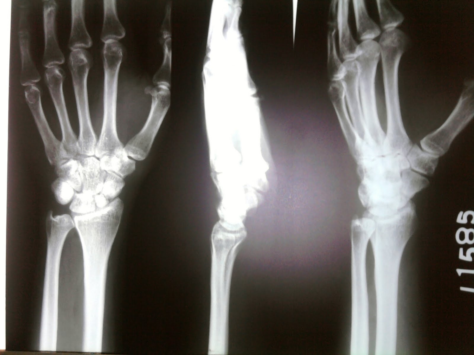
The biopsy was done 15 years ago and reported as nonspecific synovitis. Xrays- 6 degrees plantar flexion,neutral varus/valgus.


We unfortunately did not biopsy this time. As far as Sunjays comment goes. You are spot on. One cannot correct deformity with arthroscopic ankle fusion and the only failure I had with a arthroscopic ankle fusion is a deformed ankle(1/ 14). As far getting into the ankle is concerned, it is best done with ankle in dorsiflexion, no traction, direct trocar medial to lateral after entering into the capsule at the AM portal (Notch of Harty) in the coronal plane and look posteriorly to identify the joint. The clea8vage even in fibrous ankylosis is visible, if not the under x ray control using a quarter inch osteotome one could identify the same and work posteriorly with osteotome, currettes and vapr etc before fusing it. Long term secondary OA is expected when in further fusion may be needed. TER at this age is not advisable.


Bimal, 1.


The morbidity is less. The patient is home the next day comfortable. The oedema and pain post op in open lasts for a long time.


2. The union rates after scopic fusion is much higher above 95% and even the duration to fusion is less.
the xrays are uploaded for comments

Wednesday 16 September 2009

Dr. Sreenath in an aggressive mood during TKA closure.

He is now in pariyaram medical college, Kannur. Could Dr. Srenath comment on the fellowship in lakeshore hospital

.

The board room games will continue. I hope the fellowship goes on. Thanks to my fellows, juniors and colleagues we could make something of the time spent together. My teachers inspired me to share, acknowledge and gather knowledge by all legal methods. Professors like Varghese Chacko, Benjamin Joseph, NJ Mani, Bhaskaranand, Sripathi Rao, Brian O connor ,John stanley, James Richardson etc inspired us trainees. Each, added some other dimension to the attitude ie orthopedics. Team work is the name of the game. The puzzling questions raised by you guys help our degenerate neurons to fire, the addition RAM you guys add to the systems take it further. I remember a slide given to me by Jaithilak before the first Amrita arthroscopic course from the Vedas about knowledge when shared grows, cannot be divided or stolen. The system in LORC where in every member thinks to improve the system whether it is secretary, Maria school of nursing( as Bimal calls it), nurses, doctors, OT tech and all important physio adds serious value. The audits, suggestions and academic presentations push it further. Pray that the good lord continues the work where ever we are with that ATTITUDE. u know what i mean.

jake

IS THERE A ROLE FOR PELVIC SUPPORTING OSTEOTOMY


Pelvic supportive osteotomy both Shanz and Milch bachelor's osteotomy were the mainstay to avoid a trendelenberg gait in the past for paralytic dislocated hips. I understand this is still discussed in the exams and am unsure of its use even in the financially challenged.


Of late we had to convert a spate of these ostetomies to thr. Below is an example. 40 yr old male who had a shanz osteotomy 20 yrs ago, now has pian in hs Lt. hip. Neurologically normal.


1.Was a Shanz indicated?


2. Would a Chiari osteotomy been better at that time?


What now?
We did a perpendicular oseotomy at the level where the 8 mm drill from the ideal proximal entry point exited on the lateral cortex, drill the distal fragment, then sleeve and finally used a unicortical plate for additional stabilisation of the osteotomy(not always need).

Tuesday 1 September 2009

Patellofemoral instabilty in 18 yr old girl

We have a 18 yr old aspiring nurse with H/0 of recurrant dislocation patella and peripatellar pain. o/e. She has mild ligamentous laxity, No end point of MPFL with definite medial patellar laxity, +ve apprehension for patellar instabity, normal TT/TG distance, no lateral femoral condyle hyoplasia, bilateral increased femoral anteversion value- 25 degrees. She need to join college in 6 weeks. No instabilty in the opposite knee but has patellofemoral pain(lateral facet)

Do we correct anteversion first or MPFL reconstruction first or do we do both simultaneously. Do we use a IM Nail to stabilse the osteotomy? Not much of a fan of plates.

Had a comment from Dr. Sachin Tapasvi suggesting MPFL first and Big b suugesting de rotation first.

We were wondering whether to do both together. But the patient was advised conservative treatment- Physiotherapy elsewhere> Guys Magic still plays a role in medicine.

to answer sreenath, Axial cuts along the neck and trochanter superimposed on axial cuts at the epicondyle will give us the anterversion.