Sunday 25 October 2009

Infected THR with a sinus


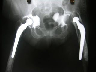
65 year old infected rheumatoid hip with femoral sinus of 4 years duration. The treating doctor told her to walk slowly till she became bed ridden with pain. Any comments


Femoral stem is jammed thru the cortical window, porotic bone and and ofcourse the mushroom shaped acetabular cement mantle.


plan


ETO, 2 stage revision, mobile spacer Plate back up in case of fracture as the bone is porotic. Vascular back up and Ilioinguinal exposure plan ready in case of iliac vessel bleed. Angio with limb movement to see if any kinking occurs. As planned we had all the problems execpt vascular injury. We used a plate to tie the ETO stabilising wires as the cortex was too thin to hold the wires. Iv antiobiotic was given for 6 weeks followed by oral rifampicin. Subcutaneous forteo was given to improve bone quality. she did not return for the second stage revision. My worries include further acetabular bone loss with abrasive wear, breakage of the spacer and rarely dislocation. May be a static spacer could avoid these problems. Sorry, I just heard she had a stage 2 revision elsewhere and is walking.

Tuesday 13 October 2009

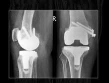
TKR in a rheumatoid valgus knee with plastic deformity of the MCL.

It was managed by a Posterior stablised knee with medial condylar (not epicondylar) osteotomy, pulled up at 30 degrees flexion to tighten the MCL. I have been lucky. Done well so far 4 years postop. May be a Contrained knee should be kept as a back up in OT

Friday 9 October 2009

TKR in a patient contralateral hip disarticulation

At the point of loading in a patient walking with a single leg with crutches and no prosthesis, the knee is in 10 to 20 degrees flexion. Possible shear vector at the poly. What would be the ideal joint repalcement. She is 60, a case of osteosarcoma 30 years post diagnosis and now ca breast in remission after chemo.

Do we do a posterior stabilized knee. LCS or a single radii knee like NRG? any comments, suggestions are welcome.
sorry she has a disarticulation of lt hip and now OA of rt. hip