Thursday 24 January 2013

Musculoskeletal Imaging has to quickly catch up

MSK imaging has improved leaps over the past  2 to 3 years. Use of 3 tesla magnets( minimum of 1.5 tesla). multichannel coils and joint specific coils for ankle. wrist and shoulder, specific sequences for cartilage have improved our diagnostic accuracy.

With insurance companies asking for MRI reports I feel it is mandatory that the imagologists update the skills and machinery to avoid leaving the poor patients troubled. I  have many times seen patients refused insurance clearance for surgery with painful conditions which need surgical intervention  due to incompetent reports and images. Oh yes, insurance  and imaging companies  are there for profit, but that should not affect the quality of care at the point of delivery.

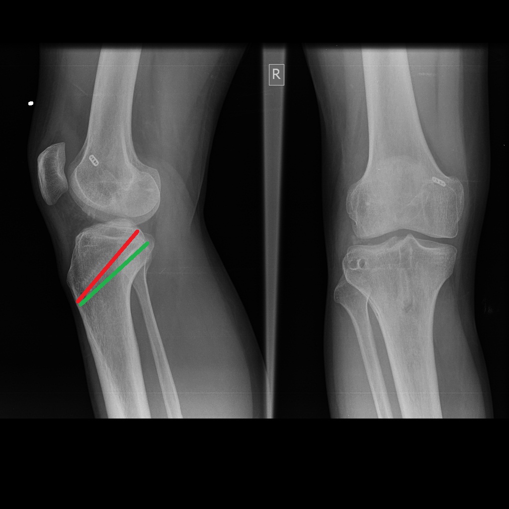
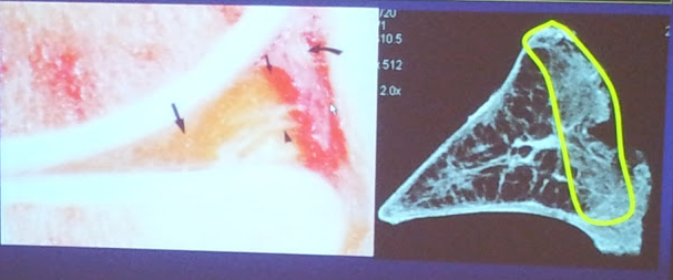
 As for us orthopods and radiologists look at the quality of pics above even differentiating the red and white zones which could help plan repair or menisectomy more accurately.  The radiologists  should quantify,qualify and  classify tears type 0 to 7 types( mesgarzadeh et al /1993.asses size and depth of cartilage lesions in addition to standard ligaments.
 Specific cartilage sequences identify chondral injury in the presence of episodic pain despite "standard normal MRI reports" and asses healing using MOCART scores to be objective about results

 I was pleasantly shocked to see some images from a 7 tesla machine.

Wednesday 23 January 2013

Tennis elbow


 The management of tennis elbow has changed over the years. The etiology is still debatable. One needs to think of D/D of  RADIAL TUNNEL SYNDROME AND RADIOCAPITELLAR SYNOVITIS which can be ruled out clinically or if needed MRI

One starts with wrist brace to immobile the extensors or counterforce brace if wrist brace can't be used. ice pacs. NSAIDs local cream and physio.

if no relief consider Inj of platelet rich plasma or steroids the choice is given to the patient as well as availability of PRP. 
At 6 months if the pain is still troubling consider an arthroscopic release of the ERCB which retracts as shown above exposing the muscle belly.The advantage over open release is that there is less likely hood of lateral collateral ligament damage. Some do repair the capsular defect which I am unsure is needed. I am impressed by the surgical results so far in recalcitrant tennis elbow

what is wrong with this pcl and plri reconstruction


 ideally the pcl tibial tunnel proximal exit has to be as shown in green above and not as in red. there is no posterior sag in this xray but patient complaints of pain on terminal flexion. he is due to review with mri

Sunday 13 January 2013

Extended trochanteric osteotomy

 
ETO believed to be a greater deliverer in revision of a well fixed stem has been a modification of wagner osteotomy which was done in the coronal plane.
 
One should measure the lenght of the osteotomy preop to get sufficeint cortical scratch fit 6 cm distal to the osteotomy. Longer the oseotomy the weaker the bone to avoid fracture. Predrilling is advisable in early experence to avoid osteotomy propagation. Curving the distal end is advised to avoid stress risers both of which I did not find necessary once good control of the saw is achieved.

Attached are some xrays where in it make efficient use of time and bone,

 to correct varus
remodelling
of the proximal femur
 easy removal of a well
fixed broken stemAdd caption

periprosthetic fracture


  A is anterior , L is lateral. The plane of ETO is the red line. Wagner is the blue line. My only modification of the ETO  is the obliquity of the osteotomy in order to increase the contact area. They are usually partial  wt bearing for 6 weeks.
 In a recent review of our cases  by Dr.Vinay,  98%  united in 6 months and all by 9 months with less than 2 mm migration

Cyclops lesion post acl recon with notch impingement

Patient presented with FFD of 10 degrees 1 years post acl recon. 
MRI revealed a cyclops lesion
Notchplasty done as above  regained his ROM. The video does not show the 4 mm burr removing the bone before    smoothening it with a shaver. Notch plasty generally is more  lateral and anterior removal of the notch. Today, this is rarely done execpt in cases of graft impingement on the anterior wall or in large graft lateral wall to prevent PCL impingement. Credits to Dr. Shibu for the edited video.

Saturday 12 January 2013

HIGH FLEXION HYPE IN TKR

 
For the past 5 years various implant manufactures and surgeons were flogging high flexion as the next best thing in TKR. The only proven study to date suggests that  highflexion ie more  135 degrees  is acheivable in patients who has high preop flexion. The implants at best protect the patient who get high flexion by increasing the contact area. Looking at the video above it is clear that the contact surface decreases as the flexion increases to the extent it could cause serious wear problems in time.  Patellofemoral pressures are another cause for concern. If the patient consistently squat, one could possibly predict wear and earlier revision.
 
Different manufactures use different methods to achieve this. Some increase the posterior condyle width( meaning remove more posterior bone which could be a problem in revision).Some change the poly curvature to achieve this. Few make the femoral box trapezoidal meaning wider box posteriorly to allow for more rotation in flexion to achieve this
 
Surgically one's options include
removal of posterior femoral and tibial osteophytes
maintaining the femoral offset with the replaced implant
Fractional lengthening of quads if quads contracture exists
increasing the flexion gap by 2 mm- potentially dangerous - could cause flexion instabilty
 

Role of arthroscopy in osteoarthosis knee

 
The indications for arthroscopy in an arthritic knee is clear cut
sudden onset sharp episodic pain in  a patient with mild arthritic pain meaning(dull chronic pain, night pain) which was well tolerated by the patient. Mcmurrys test should be positive.
 
MRI should demonstrate a meniscal tear  usually degenerative tear. MRI would also rule out ostenecrosis of femeoral condyle which again is sudden onset.
the results are even better in patients with medial compartment arthritis with sudden onset lateral meniscal tear.
 
in such cases partial menisectomy will help. the patient needs to understand that scopy will relieve only the sharp episodic pain at best and one could wash out the knee and consider viscosuplimentation or PRP injection which eve group one believes in.
 
There is no role for arthroscopic washout in an arthrirtic knee wich at best would relieve symptoms for a few weeks to months.
 please not that astenecrosi of medail femoral condyle has been reported rarely post menisectomy and should be investigated in arthritic patients with increasing pain post scopy.

Friday 11 January 2013

Drugs, dollars and doctors


http://www.scientificamerican.com/article.cfm?id=drug-research-how-we-followed-drug-research-money


Please check this article in December issue of Scientific American. The objectivity is getting lost.The influence of money colours ones views.

Monday 7 January 2013

postop cup migration in a RA osteoporotic patient

immediate postop
after postop mobilisation
This X-ray was send to me for an opinion. The initial X-rays were fine execpt for  some medialisation which in nonporotic bone would hav been fine.. On mobilising the patient complained of pain and the X-ray revealed the above.  It appears that the cup has migrated superiomedially suggesting a anterior column deficiency, but if the ilioischial line is traced up there appears to be a break sugesting posterior column breach.  Is it a T fracture.
   
First of all one needs Judet views and a ct to confirm the same. The options on table should include acetabular plates if the posterior column is compromised. A birch schnider cage if the posterior column is breached .if only the anterior column is breached one  could get away with a primary cup or a ganZ  cage and a cemented cup

Any comments or advise

 Our window for revision was 5 weeks later as the patient got chicken pox and had to postpone the revision  which increased the risks for bleeding dur to callus as well as reduced thr chances for  reduction of the T fracture  and plating. she also has a low pulmonary reserve and gross osteoporoisis. At revision we found a rotated maluniting posterior column a central/floor  defect. We elected to accept the deformity and bridge the defect with a BS cage with the inferior flange modified to a hook on table adding strength froM the tear drop( hope the flange does not break)  and cemented the cup and stem.  the stem is definitely in varus and the cementation could have been better . i do hope for a 10 yrs survival or more as she is low demand. we have started her on PTH  and VIT D.

 As a word of caution, beware of the severely porotic RA patient. using an uncemented cup is fine( if good bone stock is available) but watch out for fractures intra-op and may be postop. a cemented cup protected by a ganz cage or an antiprotrusio cage  could avoid a surgeons nightmare.

Tigth rope button stuck in the femoral tunnel


The white threads of the tight rope accidently got pulled with the button getting stuck in the  femoral tunnel(entirely my mistake) I believe. No amount of traction proximally or distally could dislodge the button as it got jammed in the femoral tunnel( check lateral view above .we got away by using a  suture button at the femoral exit. Has anyone had a similar  problem. Can the tight rope one e shortened be lengthened. . I couldn't  do anything as the button was well fixed in the cancellous bone but could not risk leaving this alone. Hence the suture button . Any comments or advise please

Tenoscopy


Synovial fold mimicking a tfcc tear

 Patient presented ith ulnar sidedpain post trauma. MRI revealed tear. scopy revealed a synovial fold mimicing a tfcc tear. removal of the fold relieved his symptoms

Sunday 6 January 2013

41 year old male with morning stiffness


The questions raised is whether there is any role for a valgus osteotomy/ hip arthroscopy.
Please comment
My views
1. adduction xray to check whether the joint space is maintained  and congruent in adduction to see the result of abduction osteotomy
2.Would A hip scopy would help map the joint cartilage to asses the wt bearing cartilage post osteotomy by assesing the cartilage in adduction as well as a chelectomy as the same time. My hip scopy experience to date is at best average
3. At present with my experience i would wait till pain develops to do a THR COC to get him > 20+ years when needed.
any other comments

failed bipolar with protrusio

 A case send for opinion. elderly person with painful hip.  Pardon the poor qulaity pics

Problems.
osteoporosis with protrusio
well fixed stem
Anterior column penetration

Solutions
1. Hip has migrated superiomedially suggesting anterior coloumn penetration- Posterior column intact hence a primary cup is possible as good posterior bone is available. CT scans could delinate if needed.( superiorlateral migration suggests posterior column penetration)
2. Dislocating the hip  from posterior  will be easier as an anterior or anteriolateral approach will make dislocation difficult- my choice will be posterior approach. Gentle dislocation with a hook. If difficult do an ETO to remove the stem and cement and use any distal loading stem
3. Would treat the patients osteoporosis too- consider PTH. Any reports on strontium in the presence of uncemented THR.
4. Would anyone consider primary cemented THR is such osteoporotic bone as  I have seen quite a few bipolar migration is severe osteoporosis any comments