Wednesday, 3 November 2021

squatting leading to dislocation in TKR

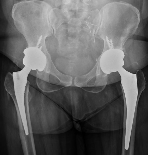
Conventional TKR is not designed for squatting. Even high flexion designs are reported to lead to earlier PF problems and wear.

Sunday, 5 September 2021

meniscal repair augmented with synovial to improve the healing in a complex degenerate tear

Menisectomy have been clearly shown to give poor long term results. Hence methods to augment and enchance repair by multiple sutures, BMAC, VGF's have been tried. we seemed to have some early good results so far with synovialaugmented repair. it is too early to be sure so we are following up the consented patients for the same.


Saturday, 4 September 2021

2 MONTH OLD DISLOCATED KNEE WITH CELLULITIS IN THE SHIN

fell 2 months after TKR and dislocated the knee. pt (BmI 35) with recurrant cellulitis on the ipsilateral tibial shin shin which is asymptomatic. esr was 110 with a crp of 20. Repeated knee aspiration was negative. incidently a gram negative UTI and large supraclavicular LN was found on admission planned for a supraclavicular biopsy, frozen section , HPE, cultures and Rotating hinge knee with arthrodesis back up. frozen section as per fieldmans criterioa showed too many PMNL in more than 5 power field, A modular titanium fusion nail and stimulan with antibiotic polymyxcin and vancomycin was inserted . Pt made to wt bear the same day evening and mobilised b haemolytic strep grew for which minimum 4 weeeks IV antibiotics is planned.

Tuesday, 24 August 2021

Greater tuberosity fracture ain’t benign

mri revealed a bankart"s lesion which was not suspected as the pt came 2 weeks post injury due to the pandemic. hence scopic bankarts reapir and suture bridge reapir of the cuff was done getting him full rom at 3 month with improving scapular rhythm. The key is do we need to look more into the tuberosity fractures for instabilty. so is mri mandatory in a tuberosity fracture to look for cuff, bankarts and SLAP tears

Sunday, 22 August 2021

Distal biceps tendon Rupture in an active persons dominant Arm

3 week old injury- Middle aged male injured rt elbow while batting sustained a bruise and swelling rt arm positive hook and squeeze tests. mri showed distal biceps tendon with retaction 8 cm above the elbow. the options of surgical treatment and conservative were discussed before repair and bracing was done distal bv class="separator" style="clear: both;">

Saturday, 3 July 2021

When will we get Sodium mri to asses proteoglycans

Wonder if the overload of neutraceuticals for joint preservation can be objectively checked by chemical MRI ie Sodium MRI


Tuesday, 22 June 2021

Greater tuberosity fractures in young adult

The incidence of dislocation of shoulder in greater tuberosity fractures are not clear.
 vice-versa is  20 percent. The chance of instability like below can be ruleout by an mri/or a good ultrasound


Saturday, 5 June 2021

MRI taken 2 months after Primary Repair of all in a clinically stable knee

the ligament appear taut  and near iso-intense


Reverse Hill sach's after anterior instabilty repair

12 months post anterior bankart  recon for a non engaging Hillsach's  Patient presented with severe pain when he woke up. MRI revealed effusion for which inflammatory markers and  aspiration was done, all of which were negative.

 3 days later he comes back with epileptic fits. The picture became clearer that he probably had a posterior dislocation with a reverse hillsachs. Immobilsed in ext rotation splint and for 4 weeks and neuro consult for his epilepsy


Monday, 31 May 2021

extensor mechanism failures post knee replacement

Causes of extensor apparutus dysfunction 
Patellar  fractures
quadriceps tendon rupture
patellar tendon rupture
patellar button loosening

Patellar  fracture
Conservative is  the main stay unless displaced  fracture or loose button

Complications of internal fixation is high
if the fracture and implant is stable - conservative
 if fracture is stable implant unstable. remove loose implant
 if fracture is unstable and implant unstable
 consider conservative first 
 Remove implant and fix
 or Recently  bypass  patella with  neoligamant and remove the implant if patella is unfixable


Thursday, 27 May 2021

Synovium - The omentum of knee

 If one can harvest synovium derived MSC to augment  meniscal healing we could find a way in avascular tears. Below is a potential technique to do the same with some early  resul


 The technique 


Involves harvest 2 cm2  synovium from the suprapatellar pouch using meniscal scissors. remove the fat and pass a looped pds or fibre wire sutures  thuogh the synovium


ts


 meniscal signs  _ve at 6 weeks


Atypical peri-prosthetic fracture due to longterm use of bisphosphonates)


 Young patient post renal transplant on long term use of oral bisphosphonates post bilateral staged THR at 2 year intervals. 


Classic features of a atypical femoral fracture on the right side and an impending fracture on the left. Medical management of the atypical fracture is still unclear.


6cm distal fixing wagner stem was used with an ETO.


Metabolic workup:


Friday, 21 May 2021

Arthroscopic Assisted Lateral Condyle elevation + Percutaneous CC Screw fixation Tibia


 Scoping  within  a few days with  low gravity flow or pressure  and using the shaver in front of the scope to suck out the clots  to see in front of scope is all always possible. Do not raise flow or pressure as it will break up th clots and compromise vision further due to bernoullis effect .scoping  3 days post injury in low velocity fall like above in 89 year old is possible.
Using cement below the graft adds to support and walking.went  PWB with a valgus unloader brace is possible for immediate mobilisation with only screws to avoid major  plates etc in patients with anticogulants etc. .

Scope and using the  acl jig allows to centralise the guide wire precisely at the depressed area and visualise elevation better than xrays specially mild tilts. A coring reamer over the guide wire within half cm below the   depressed fragment gets you a graft which u can insert under the depressed fragment after elavatio  and seal it below with bone cement or substiute


Reversed images due to introp imaging issues