Monday, 25 June 2012

18 year old adolescent footballer with Ankle pain


He has pain on dorsiflexion anteriorly  and posteriorly on plantar flexion both of which are grossly limited
options please

I am planning on both anterior and posterior scopy of ankle, removal of loose bodies and osteophytes and viscosupplimentation.  Cannot expect complete relief in view in joint space narrowing

 Post op pics today

foot and ankle problems and solutions

http://www.ankleplatform.com/.  Check this website by dr. Van Dijk. He has done a lot to popularise current methods of management of foot and ankle problems. Would recommend his course to any upcoming foot and ankle surgeon as well as general orthopod

Wednesday, 20 June 2012

Type 3 periprosthetic fracture Lt hip


 Patient a 78 year old hypertensive underwent bipolar 8 months back elsewhere and refered to my friend for mangement with a periprosthetic fracture after a fall. There is a doubt about peri-operative infection
\
Problems ansd solutions/ 
1. ? infection- Hip aspiration being done for cultures as well as leukocyte esterase test( ref Parvizi guidelines in aaos). if we get the bug even if infected would do one stage with local antiobiotic delivery of choice with HA granules or Calcium phosphate granules both of which are commercially available. 2 stage may be too much for a 78 year old.


2. Widening distal canal compromising regular cylindrical distal fixing stems unless stems locking stems like REEF or aescalup are use. I understand there is an local long stem unipolar with locking available but is a stem with no coating and is a long stem austin moore  for those who want to use it. I do not have faith in these stems.
Solutions.

Modified wagner osteotomy( Anterior) ref pic Above makes quicker sand easier operation. In this case i would prefer it to ETO. Cement and uncemented stem distally and use a bipolar head. The osteotomy helps to cement distally easily as well as remove the stem without further collateral damage. Remove the distal porocoat with a carbide burr to have better cement bone fixtion  as compared to cement implant and  hope fully avoid early loosening. ( my longest follow up for simialar case is 5 .3 years in a n 80 year old- will put it up if I can find it in my blog). If successful we could mobilise him FWB day 1 postop

3. if a high demand patient one would need to use an uncemented long HA of grit blasted stem like REEF or Aescalup stem the name of which i have forgotten

Good luck
jake

Comments by the surgeon
Hip aspiration didnot yield anything yesterday. No frank pus seen. Frozen- 10-12 neutophills. tried to dislocate first but invain. did wagner osteotomy. put in HAP granules into the distal fragment down the canal , after centrifuging it in arthrex machine with polymixin B and vancomycin . Took off the distal coating with burr. 1.2  teicoplanin was mixed with 60 gm.Unfortunately put in the cement a bit early before doughy stage  . Hence was a bit difficult to put it in.
Abt 8cm cement mantle seen in the distal fragment  from the fracture site.
Hap with antibiotics packed in at osteotomy sites and gaps proximally

I would  go PWB to FWB  as comfortable as the cement mantle is at least 8 cm  and what prevents will the pain at the fracture site.

Monday, 18 June 2012

scaphoid nonunion

35 year old rt handed office assistant fell down 3 months back. He was advised surgery by the attending orthopod but refused.  returned 3 months later with the following xray


It was planned for a percutaneous grafting and herbert screw as described the late Dr. Slade. Unfortunately the graft introducer was bigger than the proximal drill hole. The plan was to put the percutaneous wire proximal to distal Drill(red line) the proximal fragment with a larger drill (green line)to put a custom currette thru the hole and currette the cyst and bone graft the cyst with  cancellous grafts thru the custom graft introducer similar to a bone marrow aspiration needle and compress the the fracure with a herbert screw. Finally only percutaneous compression with a herbert screw was done and at 4 weeks the fracture appears to be joining.   Ideally the deformity should be corrected and graft should be used. Luckily this is uniting


Sunday, 17 June 2012

TKR iwith a bowed femur

 78 year old lady  with pain in rt thigh and knee. On Investigation her Vit D levels were low, Bonescan picked up the stress fracture. After 3 months of Vit D and PTH a tkr was planned


The limb axis films showed a 12 degree femoral bow.

Options
1. Navigated TKR
2. Custom jigs

3.What  are the options if both are not available

we did a lateral entry with the standard 5 degree femoral jig with a short rod to get the 12 degree cut.
 Navigation or custom jigs are the other options
below is the post op xray. Unfortunately the full length xrays are not availble will update it when she comes next time.

Sunday, 3 June 2012

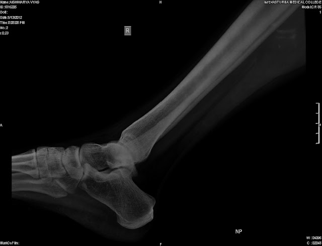
posterior ankle pain- painful os trigonum

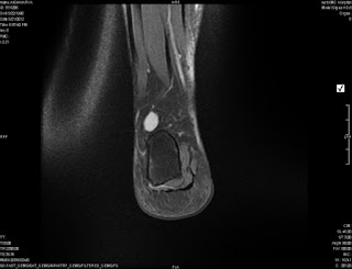
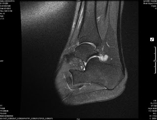
Patient presents with posterior ankle pain. Sharp Plantar flexion and external rotation causes pain . Xray confirms a large fused os trigonum and MRI shows the associated inflammation
 A local injection can be used to confirm the same.
traetment involves  hindfoot scopy and excision of both the synovitis and os trigonum

Sunday, 27 May 2012

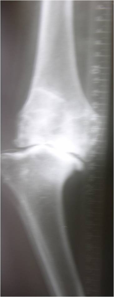
Is this the equivalent of a ball and socket knee joint

 This is an Xray of a 53 year old lady with history of septic arthritis when 15 years of age send for opinion by a good friend of mine.


This is the closet to a ball and socket knee joint. The only similar case I did was a 83 year old lady from Delhi wherein we did a Link  RHK( Pic below).  The preop pics was with valgus correction for templatingThere was no endpoint in Varus valgus under anaesthesia and the femur appeared to drop off the tibia. In view of her age we did RHK. Its 3 years now and so far successful

Here the h/o septic arthritis which is hopefully quiscent now. In view of her one could try to use a femoral  metephyseal sleeve TM may be and a Varus valgus contrained knee. AN RHK should be on table in case one tries a VV constained knee.  The baja patella is the next problem if resurfaced put a smaller button superiorly. Any other comments are welcome

Thursday, 24 May 2012

3 complex knee cases


 Below are 3 cases  send my fellow orthopods for comments. I have given my opinion and would appreciate  and welcome other contributions for this interesting cases
Case 1- 47 year old lady with pain. Apparently patient refused an arthrodesis


My views on case 1.
As  she is 47  an arthrodesis is the first option in view of the complexity, one may not get the best bearings and position. if insistent,  I would use possible a trabecular metal metaphyseal cone or sleeve with wedges to rebuild the lateral condyle. Tibia is standard wedge and stem.  Patella would be major realignment problem. The BAJA could be corrected  by using a smaller patellar button  superiorly. We do not have the skyline view to comment


The answer to bonegrafting question would be if at all it should be an entire distal femoral allograft shaped with a conical proximal( Red Triangle) end jammed into the  host metaphysis and protected with a by pass long stem( green line) as simple lateral condyle reconstruction alone is prone to resorption in the  short to medium term. Even the method I described from elsewhere is prone to fractures and resorption in the medium term. You could argue that one wants to rebulit the bone.

I have one case where in I used the sculpted femoral head( no cartilage) from amrita bone bank to rebuild the lateral condyle in a trauma(bone loss) situation. after  6 months non wt bearing  i did an arthroscopic arthrolysis and got 90 degree ROM. Tthinking about future TKR as the condyle appears vascurised on xray.  No TKR as of yet as least to my knowledge.


Below is vijays postop xray and comments ,
I used femoral metaphyseal cones and L wedges to build up the defect. Th Quadriceps was atrophic and patella was on the lateral gutter. Could mobilize by lateral release . Post op flexion is 80 now. Active extension is very weak.


Kindly opine

 Of course we need better xrays to comment on alignment. He had a RHK back up incase. The metaphyseal cone gave him a stable paltform for the femur to build on.o


Case 2 .A case of 71 yr old female having bilateral FFD of 40 degrees

 On the Lt knee in case 2  One  needs  to be careful of the MCL which,  if incompetent would need a Rotating hinge back up. I  feels if MCL is intact  I would do  try a primary knee or CCK with wedge and stem back up. Stems even if no wedges in view of the porosis and start PTH. 
At 71 RHK is techinically less demanding.

 I think the Rt side  should be standard with  just wedges and stem for tibia and a lateral femoral wedge at best. Watch out for intraop fractures and ligamentous avulsions

.Please send me the post op pics too  if possible for  all of us
 Dr mahajan comments
On Right side - I will use medial tibial wedge & for lateral femoral condyle I will use distal & posterior wedge.I will need tibial as well as femoral stems.


 The postop pics  look fine. The  rt femur is extended . It might be a stress riser if he falls. I would have stemmed  the femur too. Consider PTH injections to build bone too


I

Thursday, 17 May 2012

infected MOM bearing THR

6 years post MOM bearing presented with infected THR(psuedomonas).   The cup became vertical a year ago. Patient continued visiting various hospital and was put ion oral antibiotic. Stage 1  excision arthroplasty with HAP granules soaked in antibiotic(vancomycin) and 6 weeks of IV antiobiotics were administered
Stage 2 revision with trabecular metal augment and trabecular metal cup and wagner  stem with ceramic on poly bearing

Sunday, 6 May 2012

Osteoarthritis knee- post trauma tibia vara

 62 year old lady 5 year post open fracture tibia presently with pain and instabilty. ESR and CRP are normal. An oblique scar(red Line) below the knee healed by primary intention.


 Problems .
Medial tibial condyle has collapsed.
Mcl end point felt.
The oblique open wound distal to the tubercle is healed by primary intention and will not interfere with the  TKR incision unless osteotomy below
Sclerotic irregular eccentric canal

Options and comments please 

 Our templated plan is below including an oblique  lateral  entry to be central distally . The end result is further down. comments please.

Tuesday, 1 May 2012

Supracondylar fracture with RA and secondary OA in 65 year old lady


osteoporotic Patient with RA and supracondylar fracture
 Options
1. Fix and  TKA
2. Fix and TKA later
3. TKA now