Thursday, 30 April 2020

mucious cyst acl presenting with pain and limited flexion

Patient presented with one year of pain and restriction of terminal flexion. clinical ex. revealed stable knee with poorly localized pain on terminal flexion. No clear localising signs.. lachman1+ solid end pt. negative pivot shift. ocl and collateral stable.  no clear meniscal signs except pain on terminal flexion

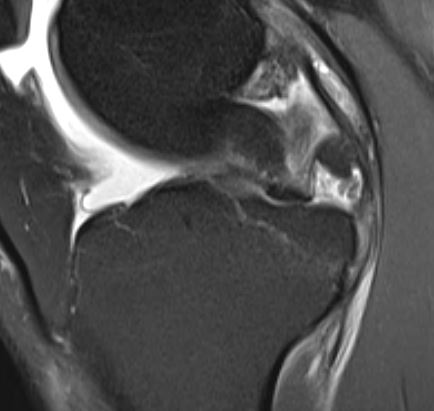
 MRI shows celery stalk appearance with cyst posterior to ACL and between ACL and PCL.
treatment
Arthro scopic cysyt decompression and intraligamentous microfracture inside the femoral attachment of ACL was done

  t

6-week MRI shows a healing ACL with the absent cyst.


6 months postop


Is ACLshaving and reconstruction necessary or the trauma of multiple small shaver punctures to decompress and cause bleeding and microfracture to release MSCs into the ligament enough at least in some cases?  The patient feels better and had full range of painless  movement 1-month post-op 


Friday, 24 April 2020

Anatomical large ceramic heads

Q

Ceramtec has finally introduced an anatomic femoral heads (decreased diameter below equator i.e. south) to avoid iliopsoas impingement. Don’t know yet about the wear characteristics to recommend usage.


Anatomic head

Thursday, 23 April 2020

1 year post micro fracture for a incidental focal lesion in in 65 year


Is  the regenerative capacity of a microfracture  in above 60 unpredictable? I had a good surprise with the  this patient?

. This was an above 60 lady with ACL and MCL injury after a fall.  the chondral lesion of about 1cm on MFC  was treated with microfracture along with stump ACl reconstruction and scopic MCL repair and orthokine intraarticular injection.  She had a Scopy for a  cyclops when the above video was taken. The defect was well filled with fibrocartilage but on palpation, it appeared stiffer  than the nieghbouring cartilage( no values) rest of her knee was pristine

Saturday, 18 April 2020


25 year male
RTA on 2/8/2019, Sustained injury to right knee
O/E:Swelling +
            Multiple abrasion
            Tenderness over joint line
            Posterior sag present
            Lachman with 2 + soft end point
            ?Medio lateral instability
            Mcmurray positive for medial and lateral    meniscus
            Varus stress test positive
            Distal pulses present


Patella inferior pole communited fracture,
ACL,PCL,LCL tear
Flipped lateral meniscus anterior horn &body tear
Medial meniscus radial tear
• inferior pole fracture was displaced and did not move in flexion x-ray hence left alone.
Right knee ACL,PCL Reconstruction + Lateral and medial meniscus (root,body) repair + LCL reconstruction done  10 days post injury


radial tear repair 


with healed radial tear pic below


He had to have an arthrolysis for a stiff knee.
ROM 20 to 70 at 2 months due to essentially lack of motivation and proper physio. the healed radial tear was visualized at 2 months during arthrolysis.. Had full ROM with a stable knee at 3 months post repair.

The meniscal healing rates are superior I feel in multi- ligament tears. 

Again a reason for early one stage repair in multi-ligament tears


ACL repair

 Over the past 4 years a trend towards repair of ACL  Sherman type 1 has gained interest. The advantages of avoiding donor site morbidity and  better proprioception are talked about. The strength of the repair with fibre wire sutures is less clear. Supplementing the repair with fibre tape augmentation has been discussed. if too tight to protect the repair the stress on the tape will invariably tear it causing IA reaction and a bit loose to avoid stress shielding by experts  defeats its purpose.
 What we clearly do is slower rehab to hopefully avoid repair failure?  Microfracture at the femoral attachment to enhance repair are being touted. 


Time will tell if we are going in the right direction.  one month post repair  mri shows a taut healing graft


Thursday, 30 January 2020

17 years post ceramic on regular poly and cemented stem in a failed Acetabular fracture

 This was the old alumina head.  ceramic head makes better sense  metal head. 
Put your money on the bearing and do a good cemented stem to avoid one more failure mode at the taper

scope assisted tibial plataeu fracture fixation in a 90 year old

Using an acl jig the depression is elevated and stabilised with raft screws. bone cement was used to support and fill the tunnel and mobilised day one with an unloader brace 


complex varus knee- posterior medial subluxed knee

 65 year old RA knee with subluxed rt knee. 

Although the initial x-ray looks like a posteriormedailly dislocated knee. tmanual reduction and xray and stress shows intact LCL and MCL making the surgery simple with standard PS and tibial wedge and stem

Stress xrays


Thursday, 12 December 2019

Bony root injuries to meniscus


SUSTAINED ACL WITH ? PCL AND LCL INJURY
 EUA REVELAED ONLY ACL, PCL Grade 1 WITH INTACT ACL
 MRI REVEALED POSSIBLE ROOT AVULSION LATERAL  MENISCUS MORE THAN MEDIAL. SCOPY REVEALED LATERAL MENISCAL ROOT WHICH WAS UNSTABLE HENCE A SCREW AND ROOT SUTURES THRU AN ABS BUTTON WAS DONE


lllr larger root g\fragmnt with PCL fixed with 2 screws and percutaneous scope assisted mcl repair


Friday, 22 November 2019

aseptic loosening hip


 47 year old female 6 years post primary thr with progressive pain. The acetabular cup appears to beloose and migrated  superior- laterally with significant posterior column bone loss. got away with a jumbocup and autologous grafting. host bone contact was just 60 percent. the below in a 11 post op rev pics 

dejour type 2 trochles with patellofemoral instabilty


Presented with pain and swelling lt knee. rt knee had a patelelofemoral mpfl reconstruction 10 months ago. no major complaints, although apprehension is positive, post trochleoplasty and MPFL reconstruction pics are below


Trochlear bump ie: ski jump has be removed the trochlear groove lateraised correcting the TT/TG distance , trochleoplasty and mpfl  reconstruction done


Thursday, 22 August 2019

Tkr in Arthritic knee with tibial fracture

51
51 year old Pt 10 years post patellectomy presented with fracture  Tibia. Patellectomy was  for infection post fixation for a fracture patella> scar had healed by secondary intention


After couselling the patient we did fixation and Tkr with a stemmed Tina and mobilesed non wt bearing for six weeks
Achieved ROM 10 to 80 degrees with and extensor lag of 10 degrees at 2 months
She was glad to get back to work at 2 months


The option of 2 stage was advised elsewhere is acceptable but the time between 2 stages and overall rehab may be close to 6 months


Friday, 22 February 2019

MOM lt hip with rising cobalt Levels

71  yr old patient 12 yrs after lt THR  with rising cobalt levels  of one year duration with no local symptoms. Netflix movie "Bleeding heart"  convinced the patient of revision inspite of lack of symptoms.  All other blood parameters. Bone scan and MARS MRI done in 1.5 T machine was normal.

 There was local metallosis or infection.  manage d to retain a well fixed sleeve and changed the cup to 56 from 54 and a lateralised 4+4 Calcar replacement stem with ceramic on poly THR was done

 This was my revision of Srom in 3 cases where in I could retain the sleeve and patient walked like a primary hip.
 the rt side has a COC revision hip done after the MOM fiasco