Sunday, 29 April 2012

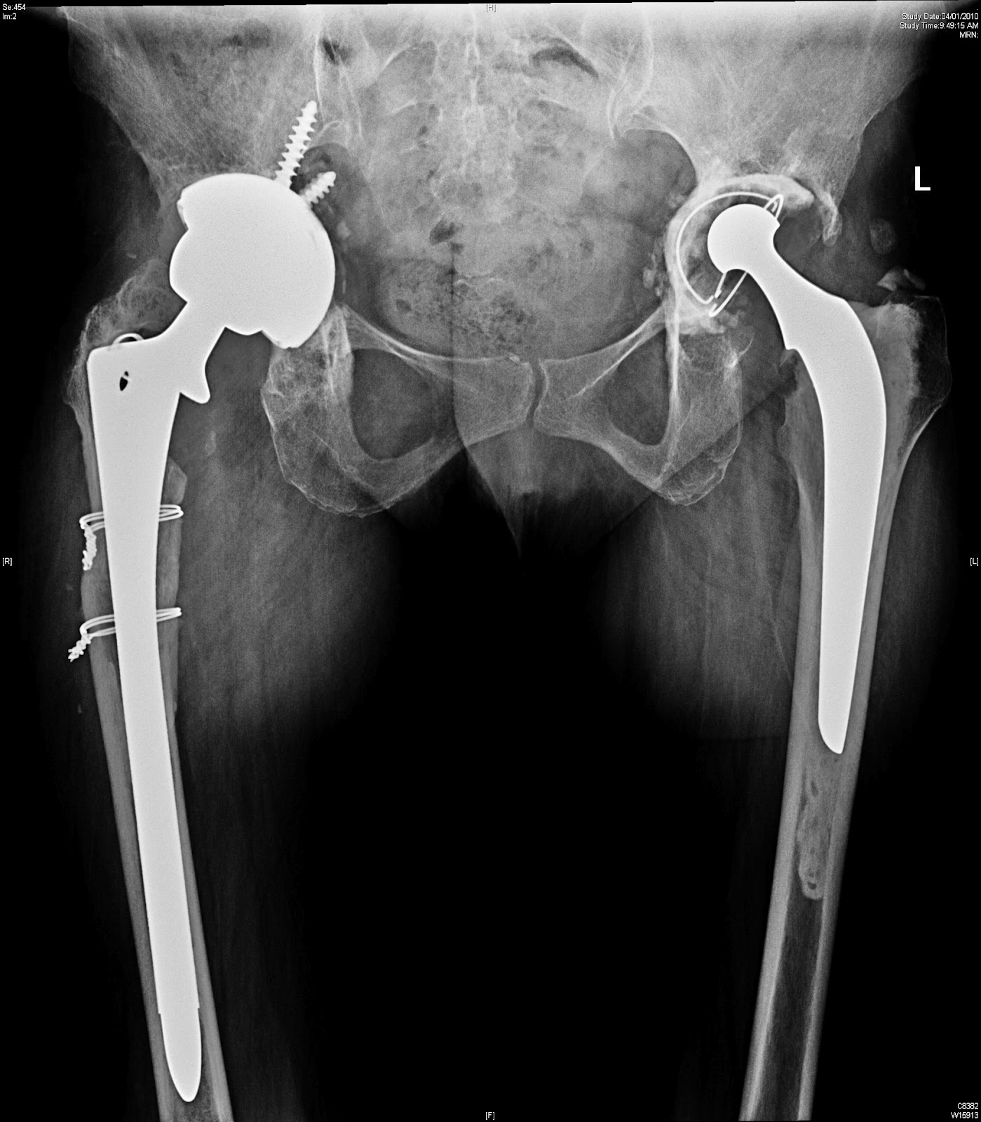
Bilateral hip ankylosis with HO

 Case refered by Dr. Mukesh Ladda from nagpur for opinion.
Please  give your views and comments

54yr male diabetic presented with bilateral stiff hip since one yr,slowly progressive to total stiffness since last six months.
History of septicemia with multiorgan failure one n half yr ago,recovered well but unable to stand or sit due to bilateral stiff hip.

His  question are as follows? My comments below are open to your valuable comments and views
1.Is THR only option?
With the sort of long standing Ankylosis the articular cartilage nutrition suffers and result in fibrous or bony ankyloisis. The question whether this was the result  of septic arthritis or due to neuraxial injury could be partly answered by ESR, CRP and intraop frozen section. Being a diabetic an HBAIC is advised. IF proven to be infected could consider girdlestone with local antibiotic delivery could be a choice before THR. If not A THR with the largest possible head to avoid dislocation

2.What will be the status of Abductors after removing HO?
 I would go dead lateral, split the TFL with anterior and posterior with bone slivers from the G. Troch. Do a neck ostetomy and remove the head, Ream or piece meal. After this the bone anterior and posterior can be removed  slowly exposing the bone subperiosteally and reove all the impinging bone. You want to watch for N/V damage  palpating vessels and preop MRI angio  if needed to asses where the vessels are embedded if needed. The all important homan's retractor, electroquatery  helps exposing the HO. The one similar case i did had a compartmental syndrome of thigh and femoral neurpraxia which recovered partially in 6 months. The contralateral hip was done 6 months later and laser was used toquarterise the bleeding bone to avoid compartmental syndrome. Those days we did not have any local thrombin delivery to stop bleeding if possible.

3.When should i do the second hip(gap between two THR)?
I would wait 3 to 6 months only for recovery
4.What are  the chances of recurrence? In my only experience of a bilateral case the xrays below the hip failed with a psuedo tumor due to MOM articulation with an XL head.

5.Radiothearphy protocol pre & post op?
My choice is radiation the morning before operation( experimental evidence in literature)

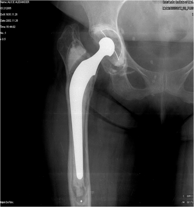
Below is a similar case I discussed before, So far I could trace only the preop and one post op at 3 months. will trace the entire series  in a few days once I get back to get the whole story.

Dr. Mojieb's Comments are added below

1- Would pre operative aspiration add value too to ascertain the possibility if this being post sepsis or not
2- Any role for Posterior Approach ???
That might give u a better opportunity of visualizing & protecting the hip abductors.The only issue will be, where to osteotomize the ankylosed  hip which I think u can do it a bit low , just above the lesser trochanter , this will help you retract the whole femur + abductors out of your way than ream away the acetabulum piece meal initially than as the landmark for the ant+ post columns become available u can place the Homan.

I like your idea about the neurovascular structure concern & the use if laser for bone cauterization
I personally have not used laser before but seems to be a great idea.

Sunday, 22 April 2012

Asymmetric polywear in a tkr

I do not have the initial postop xrays taken at the time of index procedure done elsewhere. Hence, not sure whether improper balancing was one of the causes for  early  polywear ( 7 years)


 This was revised with legion revision system. The implants werte well fixed but the poly liner is not available anymore at least in india( FS knee) hence the total revision

Tuesday, 17 April 2012

lessons learned-Sequelae of open fracture talus with medial malleolus fracture


He has  pain on wt bearing with Varus hind foot and gross painful restriction of ankle movement. Varus of ankle can be partially corrected short of neutral.  Subtalar movemnts are terminally restricted but painless. Midfoot and tendons are normal.


1. Is this AVN with collapse.
2. Malunion with Sec OA
3. Investigations if any

4. what are the options
 we did an arthroscopic ankle fusion. the reasons and problems are discussed in the comments


Now after 3 months the ankle is solid fused has lateral pain from an abuting osteophyte or should one call it unionophyte. The disheartening xray is of .> 10 gegree varus which i feel i would  need correction thru a transfibular approach when symptoms arise. He is on a rocker heel slippers to simulate heel toe gait.


1.      He was referred for ankle replacement. We did a bone scan to rule out AVN and decided to do an arthroscopic ankle fusion as he was young and works as a electrician. I believe an ankle replacement at this age may not be a good idea. As his subtalar joints were relatively normal an ankle fusion was chosen. In view of his varus deformity we planned to open if the deformity was not corrected, By clearing the lateral gutter with Shaver and burr we could correct the varus and hence only the medial malleous screws were removed and percutaneous tibiotalar screws were inserted. Unfortunately one of the guide wires broke and intraop screening showed it to be intraosseous. However the postop xray showed the wire protruding into the sinus tarsi. I am hoping for an early fusion and hopefully no subtalar symptoms


Lessons learned.


1. Do not attempt arthroscopic fusion in the presence of varus deformity which is not passively correctable

2.My reason to attempt the same was due to the medial and lateral as well as anterior skin inscisions which would compromise wound healing as well as after taking out the lateral osteophyte i felt i corrcted the deformity refer xrays above which I know now was never a true AP

The Xrays below are refusion at  6 weeks postop. The fusion clinically feels solid. We have delayed wt bearing and left the foot unsupported inspite of bridging anteriorly and posteriorly as open fusions generally take 3 months to fuse.

Over all arthroscopic ankle fusions have above 95% fusion rates at 12 weeks. My personal series of 18 cases have all united execpt for 2 malunions both of which the indications were flawed due preop varus deformity as above which were only partilly corrected.


Sunday, 15 April 2012

Price one pays for delayed revision in aseptic looseningr


This patient, a 44 year old rheumatoid lady underwent revision of rt hip in 2005  with MOM where in  we  advised revision of the lt. cup. Financial reasons or otherwise, she comes  7 years later with the cup migration. comments please. Her metal ions are fine. The xrays are in chronological order. Early revision could have saved much bone and made surgery easier.
1 
Would have loved to revise early without bone loss. Superiomedial migration suugest anterior column deAs the anterior column was compromised we could get good host bone with more than 70% contact with a jumbo trabecular cup and a wagner stem 

Monday, 6 February 2012

modified hardinge approach

Modified hardinge Approach maintaing 2/3rds of the medius  and releasing towards the lesser trochanter and not vertically downwards maintain the medius- lateralis tension band as well as exposes the acetabulum and the femoral shaft necessarsary for standard THR. Why violate tissue when you dont need to?

Wednesday, 1 February 2012

Revision ACL

5 months after ACL reconstruction Rom 15 to 90. why should this happen. What  to do now


On Scopy a "cyclops" lesion was seen. this was shaved of and a wide notch plasty was done to prevent further impingement. Full extension was acheieved however flexion was still limited. The suprapatellar pouch and the medial and lateral gutters were leared of the scar. Patellar mobility and full rom was achieved with medial and lateral patellar release which was the next stage of the plan.

Patellar impingement with femoral condylar plate

28 year old Salesman 8 month post orif of Intercondyalr fracture femur. Presented with ROM 10 to 80 degrees ans pain on extension.
O/E. decreased patellar mobility, Clicking band(Plica) lateral to patella at 20 to 10 degrees of extension. Cruciates and collateral stable.  views please

Monday, 19 December 2011

aseptic lossening of cup in a 62 year old female 4 year postop

Resonably fixed charnley stem and loose cup


Why did it fail?
Poor acetabular cementing
Medial wall penentration  could cause other problems but not loosening.
Tips for good acetabular cementing
Dont ream the subchondral bone off
The last reamer used should be at least 2 mm or 4 mm more than the cup size to get at least a 2 mm cement mantle with good penetrarion
Don't ream the subchondral bone. Multiple(8 to 10 in no) 4 mm peg holes better than large key holes. Make the key holes wider than the  mouth for macro locking.
Dry acetabular bed before cementing
Pressurise after inserting doughy cement
Apply cement on the back of the cup too to avoid blood at the cement implant interface
Insert the cup locking the inferior cement first in an open position before  bringing it to 40 degrees and anterverting. Maintain pressure with the head shaped pusher while removing excess cement from the periphery till its hard
Remove any peripheral osteopyhtes to prevent impingement
 Finally it is easier to do an uncemented cup well than a cemented cup for the beginner
 Do we revise the cup alone?
If we do this, the dislocation chances are higher due to neck cup impingement. To correct this one needs to revise the stem to use a 32 mm or 36 mm head.
 How do we revise the stem?
a. ETO and distal fixing uncemented stem
b. Cement on cement revision as advised by  Dr. Phil Roberts
 We did a cement on cement stem and an uncemented cup without removing the medial cement, obtained could posterior superior contact
Used a high speed drill and stem extraction and cementeda tapered stem with an oxinium on xlpe liner

Sunday, 4 December 2011

52 year old male with aseptic loosening at 3 years

The patient was advised by the surgeon that the implant was rejected.. This is an example of bad cementing or polywear causing loosening.

Removal of debri, washing out the blood, dry field are mandatory before cement application. Pressuring, cement gun introduction and timing of implant introduction depends on the type of cement and room temp.
This was definitely not rejected
jacob

Sunday, 20 November 2011

migrated cup

Acetabular loosening are best managed early. Do we need to wait till major bone loss before revision? This is a 55 year rheumatoid lady 10 years post primary hip replacement.
Problms
Eplantation of cup
Vscular risk
Acetabular defect managnement
well fixed stem

Saturday, 19 November 2011

Knee pain


Sports were designed to improve fitness of ordinary mortals. In our quest for higher level of fitness we sometimes damage joints beyond repair. Some amount of cyclical loading is good for cartilage (the softer shock absorbing ends of bones) nutrition. A Swedish prospective study comparing degenerative wear with exercise compared daily exercise, with thrice daily exercise and no exercise and concluded that thrice daily exercise probably had the least wear.

Eg. Cartlilage culture

Once a mechanical change to the alignment or stability of knee occurs, it becomes the beginning of the end of knee. Ligament injuries which contribute to stability are akin to suspension and wheel alignment of our cars. Your continuous driving without correction wears out the tyres similar to the softer cartilage of the knee.

Degemeric MRI
Pain is a protective phenomenon evolved to prevent further damage to oneself and administering of pain killers alone without investigation and treatment of the cause could further deteriorate the joint.. As far diagnosis of cartilage injuries, MRI scans upto 3 Tesla magnets with de-gemmeric software to look at cartilage has improved the diagnosis of cartilage injuries. These days easy reproducible minimal access surgery is available world wide to correct alignment, ligament reconstruction and resurfacing of cartilage injuries using osteoarticular grafts, bioscapholds and matrix assisted autologous implantation.

The use or abuse of exercise is ever increasing in our quest for healthier hearts. Over doing the same has exacerbated the possible incidence of osteoarthritis in knees. Weight reduction is the first simple step to reduce the wear across the cartilage. Six times one’s the body weight goes through our knees while walking. It is akin to driving a truck with cycle tyres. The smaller bony part of the knee has to take the excessive load which is already getting stiffer due to chemical changes and decrease water content with aging. Stronger and fitter muscles do take part of the load.

The role of neutrocaeuticals for arthritis has been controversial. Glucosamine and chondroitin sulphate are normal constituents of articular cartilage, but only 8% gets absorbed and how much reaches the knee is unclear. There are no studies to prove that the cartilage becomes normal with ingestion of neutricaeuticals. It at best could reduce the rate of degradation and might have analgesic effects. There use has been widespread so as to avoid the deleterious side effects of long term use of analgesics which are proven to cause Gastrointestinal and renal problems.

Viscosupplementation have been used to delay the inevitable and recently been talked about as a disease modifying intervention. This is yet to be proved and at best would be a stop gap arrangement till the inevitable knee replacement occurs.
Knee replacement has been shows to be in medium to long term, the best option once end stage arthritis occurs. Newer designs, materials like oxinium and crosslinking of polyethylene have proven to reduce the wear rates and improve the longevity of the implant. The complications of Pulmonary embolism, infection has been decreased with better drugs and early mobilization. As younger arthritic patients are demanding better function newer partial knees are being tested.


Correction of alignement(HTO) in an young patient with arthritis


preop with knee deformity


post knee repalcement op of the same patient

Pre Knee replacement

post knee replacement

Reducing blood loss during joint replacement

Methods to avoid transfusion, more so in bilateral TKR are possibly being achieved by using transxemic acid ,Thrombin, aquamantis and other methods. Our preliminary trial shows a trend towards minimising blood loss in Rheumatoids and bilateral tkrs using FLOSEAL.

Does size matter

The size of the incision or the implant does nothing to the longeivity or function of the implants. It is the technique, quality of bearing, fixation and alignment which is paramount. The approach whether anterior or posterior if done well does not matter. Why are we stuck on the size? yes, it could be a marketing tool. These fads fade away  and what matters most is the function and longievity. Smaller Proximal loading stems might have a role in preserving proximal bone. Till date No hip other than resurfacing maitain bone in gruen zone 1 and 6. With the metal on metal controversies surface replacement is used in select cases only.

Wounds heal side to side and possibly better rehab and multimodal analgesis will help to get the patient up quickly.

Thursday, 17 November 2011

migrating stem

65 year old lady 10 years post primary THR.


Well fixed cup with no major wear.


Migrated stem with varus remodelling of proximal femur

Plan- ETO for correcting the femoral deformity while revison. cup was also revised . unfortunately only 22 mm heads were available in india then in 2000.


type 3 valgus knee


76 year old lady with 60 degree valgus with no medial end point
Only A rotating hinge TKR is possible in heras she has an incompetent MCL.

Risks include a foot drop postop when you correct the deformity.


I used the Link Devise succesfully in her. She is 3 years postop with no major problems