Patient with anterior pain on dorsiflexion exacerbated with anterior joint line presssue. Anteriomedial oblique xrays show the impinging osteophyte
Saturday, 27 October 2012

Thursday, 11 October 2012
arthroscopic management of meniscal root avulsion
Saturday, 1 September 2012
Failed ACL reconstruction


Any takers on management and technique
My options given to the patient.
1. Scope remove the intra articular screw ASAP and do a revision ACL. However I warned him that in case of severe OA one might consider not doing an ACL. One could envsage problems only with the tibial tunnel screw. I plan to the tibial tunnel entry more medial exiting at ht lateral half of the medial tibail spine. Femoral tunnel isnt a problem as the previous tunnel is vertical.
Post op pics are below. The screw had damaged the medail edge of the lat femoral condyle adjacent to the notch( Like a wide notch plasty)


Wednesday, 29 August 2012
post acetabular fracture - post milch batchelor osteotomy
Case for opinion-
This 52yr old gentleman had girdlestone followed by shanz ostetotomy in 1995 for neglected fracture dislocation left hip.
Now he complains pain around same hip and wants THR
Bloods are normal
The senders plan is for uncmented Depuy ceramic on poly with S-ROM stem.
Will he need Subtrochantric shortening Ostetotomy?


This 52yr old gentleman had girdlestone followed by shanz ostetotomy in 1995 for neglected fracture dislocation left hip.
Now he complains pain around same hip and wants THR
Bloods are normal
The senders plan is for uncmented Depuy ceramic on poly with S-ROM stem.
Will he need Subtrochantric shortening Ostetotomy?

My view is
1. I need judet view/CTscan to see the acetabulum the posterior colum and wall in particular
2. intraop specimens for frozen section and culture
3.Looking at the AP a primary acetabular cup is possible.
4. I wouldnt do an SROM as in a similar case the corrective subtroch osteotomy went in for nonunion as SROM is essentially a distal fitting stem and the hold on the distal fragment is not rigid as a distal loading stem like Solution or Echelon

5. You wouldnt need a shortening most likely but would correct the deformity where the central axis of the proximal fragment(Red Line) reaches the medial shaft- an opening variety(ORANGE IS THE LINE OF THE OSTEOTOMY PERPENDICULAR TO THE RED LINE) and ream and hold the distal fragment tight with solution or echelon COCR stems . graft the result defect with the cancellous graft> plrase strip the v lateralis only at the osteotomy keeping the muscle through a vertical split maintaing the the extramedullary blood supply of the distal fragment.
Sunday, 19 August 2012
TKR in postraumatic stiff painful knee
63 yearold lady with post traumatic arthritis- 5 years post open type 2 fracture tibia
treated primarly by illizaro and screw presented with pain and 0 10 to 30 degress rom. No sinuses. ESR and CRP normal.. Multiple scars on the proximal tibia as sequelae of open fracture. Never had sinuses postop. Piperacillin- tazobact with CAPO4 pellets were introduced into tibial canal to combat bugs if any and a fractional lengthening of quads was done.to obtain 100 degrees flexion.


We used an offset stem. Long stem was avoided so as to avoid further osteotomy inscisions as well as to avoid exposing the luscent ares of tibia.
Introp frosen section and leucocyte esterase tests were negative.


Tuesday, 14 August 2012
33 yer old male post osteotomy for opinion from dr. rajesh

Ensure that the pain is from the hip itself as the lateral joint space is reasonable although inferiomedially it looks narrower. If in doubt put local in the hip and reasses pain relief.
ANY takers for FAI here- SCOPE and proceed
If a THR is planned 1would consider the follow Problems including insitu implant- remove the scews after dislocation to avoid intraop fractures. the plate can be left alone. sometimes one might be able to put a small stem between the screws in cemented situation. Send intraop cultures
the stem should ideally be longer then the plate to avoid postop fractures.
some doctors use preop SWD to heat the implant to loosen it preop, other heat the screws with the cautery to loosen the screws.
keep broken screw removal kit in case of problems and include the philips head screw driver incase of older generation screws
2. Varus neck with a shorter neck. bigger offset stems would help to maintain the abductor tension as well as to prevent lengthening in case of using a hard on hard bearing obtaining a tight reduction and avoid stripe wear.
longer stem preferbly modular proximal loading stem like SROM to make revisions easier in case of ceramic head fracture. one could use a longer cemented sem like exeter with offset options.
3. avoid introp and postop fractures.
as far as your questions on entry point to avoid varus stem.

I would extend a line proximally on the xray as shown below from the medial edge of lateral cortex of the diaphysis and measure it from the lateral trochanteric cortex(orange Line) to make it your starting point introp. some companies have a lateraliser instrument to further lateralise the entry.
good luck
Thursday, 26 July 2012
Ossron(cultured osteoblast) in AVN hip
Above is the xray of a 28 year doctor with pain in lt hip of 3 months duration. The mri showed a gr 2 lesion in lt hip and grade 1c rt hip. We attempted acteocyte culture and at 5 weeks the postop xrays are below. We are awaiting MRI pics to look at revascualrisation. His symptoms are better but that could be due to decompression alone.
pleasantly surprised


Wednesday, 18 July 2012
Complex Hip- OA with intertroch nonunion with implant in situ
Any takers for this complex hip pics send to me by my good friend


Charan whats the problem now and solutions
as expected the hip dislocated

The options now
The cup appears too anteverted as well as vertical. . with a flying trochanter adding to the problem.
my advise is to revise the cup. use larger head if possible and use a trochanteric cable plate.if the troch is too soft to hold wire over a titanim mesh. if this does not work only a capture cup or a tripolar head is the only solution.
My friend revised the cup and stabilised the hip. his trochanter has dissapeared. In this situation anchor the glutea to the reamaning bone or if that is not possible anchor the two flaps of TFL to bone leaving the distal portion free to inset distally. Abductor bace for 6 weeks

Tuesday, 17 July 2012
11 years post Single bundle PCl reconstruction

o/e.
PCL was close to gr 2 solid end pt. no meniscal signs or joint tenderness Dial test negative. Xrays show minimal medial compartment narrowing as compared to the normal side. No patellofemoral changes.
Inspite of old technique the PCL recon was worth in a young man. I believe the present day literature support even in isolated PCL injuries more than grade 2.


My
Monday, 25 June 2012
18 year old adolescent footballer with Ankle pain
He has pain on dorsiflexion anteriorly and posteriorly on plantar flexion both of which are grossly limited

options please
I am planning on both anterior and posterior scopy of ankle, removal of loose bodies and osteophytes and viscosupplimentation. Cannot expect complete relief in view in joint space narrowing
Post op pics today
Post op pics today


foot and ankle problems and solutions
http://www.ankleplatform.com/. Check this website by dr. Van Dijk. He has done a lot to popularise current methods of management of foot and ankle problems. Would recommend his course to any upcoming foot and ankle surgeon as well as general orthopod
Wednesday, 20 June 2012
Type 3 periprosthetic fracture Lt hip

\

1. ? infection- Hip aspiration being done for cultures as well as leukocyte esterase test( ref Parvizi guidelines in aaos). if we get the bug even if infected would do one stage with local antiobiotic delivery of choice with HA granules or Calcium phosphate granules both of which are commercially available. 2 stage may be too much for a 78 year old.
2. Widening distal canal compromising regular cylindrical distal fixing stems unless stems locking stems like REEF or aescalup are use. I understand there is an local long stem unipolar with locking available but is a stem with no coating and is a long stem austin moore for those who want to use it. I do not have faith in these stems.
Solutions.


3. if a high demand patient one would need to use an uncemented long HA of grit blasted stem like REEF or Aescalup stem the name of which i have forgotten
Good luck
jake
Comments by the surgeon
Hip aspiration didnot yield anything yesterday. No frank pus seen.
Frozen- 10-12 neutophills. tried to dislocate first but invain. did
wagner osteotomy. put in HAP granules into the distal fragment down the
canal , after centrifuging it in arthrex machine with polymixin B and
vancomycin . Took off the distal coating with burr. 1.2 teicoplanin was
mixed with 60 gm.Unfortunately put in the cement a bit early before
doughy stage . Hence was a bit difficult to put it in.
Abt 8cm cement mantle seen in the distal fragment from the fracture site.
Hap with antibiotics packed in at osteotomy sites and gaps proximally
.jpg)

I would go PWB to FWB as comfortable as the cement mantle is at least 8 cm and what prevents will the pain at the fracture site.
Monday, 18 June 2012
scaphoid nonunion
35 year old rt handed office assistant fell down 3 months back. He was advised surgery by the attending orthopod but refused. returned 3 months later with the following xray


It was planned for a percutaneous grafting and herbert screw as described the late Dr. Slade. Unfortunately the graft introducer was bigger than the proximal drill hole. The plan was to put the percutaneous wire proximal to distal Drill(red line) the proximal fragment with a larger drill (green line)to put a custom currette thru the hole and currette the cyst and bone graft the cyst with cancellous grafts thru the custom graft introducer similar to a bone marrow aspiration needle and compress the the fracure with a herbert screw. Finally only percutaneous compression with a herbert screw was done and at 4 weeks the fracture appears to be joining. Ideally the deformity should be corrected and graft should be used. Luckily this is uniting


Sunday, 17 June 2012
TKR iwith a bowed femur
78 year old lady with pain in rt thigh and knee. On Investigation her Vit D levels were low, Bonescan picked up the stress fracture. After 3 months of Vit D and PTH a tkr was planned


Options
1. Navigated TKR
2. Custom jigs

3.What are the options if both are not available
we did a lateral entry with the standard 5 degree femoral jig with a short rod to get the 12 degree cut.Navigation or custom jigs are the other options
below is the post op xray. Unfortunately the full length xrays are not availble will update it when she comes next time.
Sunday, 3 June 2012
posterior ankle pain- painful os trigonum
Patient presents with posterior ankle pain. Sharp Plantar flexion and external rotation causes pain . Xray confirms a large fused os trigonum and MRI shows the associated inflammation
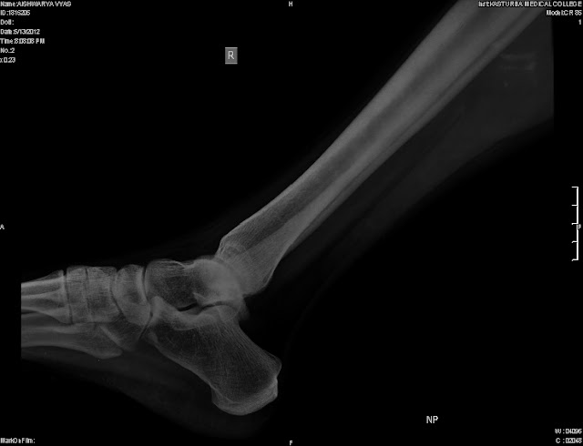
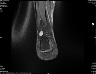
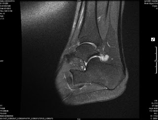 A local injection can be used to confirm the same.
traetment involves hindfoot scopy and excision of both the synovitis and os trigonum
traetment involves hindfoot scopy and excision of both the synovitis and os trigonum
Subscribe to:
Posts (Atom)